Home Articles Glaucoma: evaluating retinal protection in preclinical models

Glaucoma: evaluating retinal protection in preclinical models

Glaucoma is one of the main causes of visual impairment and blindness worldwide. Protecting retinal cells is a potential therapeutic strategy to limit vision loss, making preclinical models important tools for investigating retinal injury and evaluating new approaches to treatment.

Table of Contents

VectorB2B combines experience in basic and translational Vision Sciences research with established in vitro and in vivo glaucoma models. These capabilities are supported by techniques for assessing retinal structure and function, allowing researchers to examine the effects of potential therapeutic interventions.

Glaucoma research: assessing retinal injury and therapeutic response

Evaluating the therapeutic potential of a compound in glaucoma research requires preclinical models that reproduce key features of the disease and allow the effects of potential treatments on retinal structures and function to be investigated. One of the central questions is whether an intervention can protect retinal tissue following injury, providing evidence to support further investigation.

This evaluation can draw on complementary findings from in vivo models. Assessments of retinal structure, electrical activity and visual function provide different perspectives on the effects of retinal injury. Combined with appropriate experimental models, these techniques support the investigation of protective responses to potential therapeutic interventions.


Preclinical glaucoma models at VectorB2B

VectorB2B has established two experimental approaches for glaucoma research, based on elevated hydrostatic pressure in vitro and transiently increased intraocular pressure in vivo. These models provide different experimental settings for investigating retinal injury and evaluating potential therapeutic strategies.

In vitro models

The in vitro model uses an elevated hydrostatic pressure (EHP) chamber, allowing the effects of increased pressure to be investigated under controlled experimental conditions. This approach forms part of VectorB2B’s portfolio of experimental models for studying processes relevant to glaucoma.

In vivo models

VectorB2B portfolio includes a well-established in vivo model based on transient elevation of intraocular pressure, which induces retinal ischemia-reperfusion (I/R) injury. This experimental approach allows researchers to assess retinal damage and investigate responses to potential therapeutic interventions.

The I/R model provides a platform to investigate new therapeutic approaches and has been used to evaluate the protective effects of adenosine receptors modulation, including A2A receptor antagonism with istradefylline (KW6002) and A3 receptor activation, as described below.


Retinal structure and visual function assessment

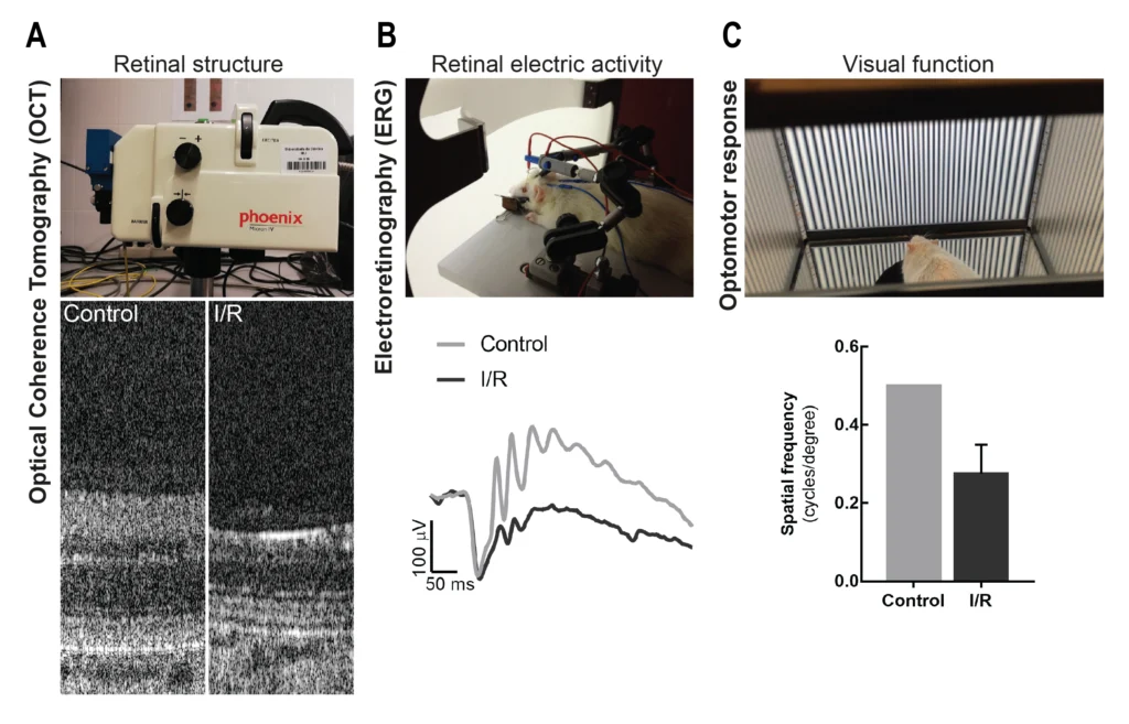
In preclinical glaucoma research, assessing retinal injury involves examining changes in retinal structure and function. Several techniques with clinical relevance can support these evaluations in in vivo models:

  • Optical coherence tomography (OCT): assessment of retinal structure;
  • Electroretinography (ERG): evaluation of retinal electrical activity;
  • Optomotor response: assessment of visual function.

Figure 1 illustrates the application of these techniques, including comparisons between control animals and those subjected to I/R injury.

The OCT images show alterations in retinal structure, while the electroretinography recordings illustrate differences in retinal electrical activity. Optomotor response measurements provide an additional perspective on the effects of retinal injury on visual function.

How preclinical models support glaucoma research and the evalua-tion of retinal protection
Figure 1: Assessment of retinal structure, retinal function and visual performance following ischemia-reperfusion (I/R) injury. (A) Representative optical coherence tomography (OCT) images, (B) electroretinography (ERG) recordings and (C) optomotor response (OMR) measurements comparing I/R-injured animals with control animals.


Istradefylline (KW6002): evaluating retinal protection in vivo

Istradefylline (KW6002) is a selective antagonist of the adenosine A2A receptor (A2AR). The compound was approved as an adjunctive treatment for Parkinson’s disease in Japan in 2013 and by the US Food and Drug Administration (FDA) in 2019.

Its potential for retinal protection was investigated using an animal model of transiently elevated intraocular pressure. Following the induction of retinal ischemia-reperfusion injury, istradefylline was administered orally to assess its effects on retinal damage.

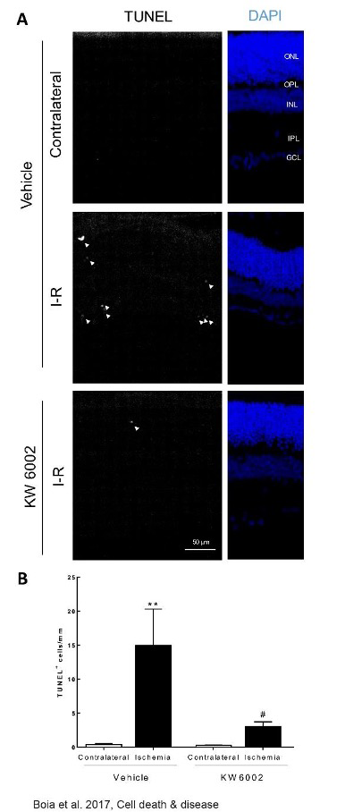
The results demonstrated a protective effect against retinal injury. Figure 2 presents retinal sections analysed using TUNEL and DAPI staining, alongside the quantification of TUNEL-positive cells. In animals subjected to ischemia-reperfusion injury, an increase in TUNEL-positive cells was observed compared with contralateral controls. Treatment with KW6002 reduced the number of these cells, providing evidence of retinal protection in this experimental model.

How preclinical models support glaucoma research and the evalua-tion of retinal protection
Figure 2: Effects of orally administered istradefylline (KW6002) on retinal cell death in an I/R model induced by transient elevation of intraocular pressure. KW6002 (3 mg/kg) was administered by oral gavage 2 h post injury and until the end of experiment (7 days of reperfusion). (a) Cell death was assayed in retinal cryosections by TUNEL assay at 7 days of reperfusion. Nuclei were stained with DAPI (blue) and representative images are depicted. (b) TUNEL+ cells (gray, some TUNEL+ cells are indicated with arrowheads) were counted and expressed per mm of retina. **P<0.01, significantly different from contralateral eye; # P<0.05, significantly different from I-R retinas of KW6002-treated animals, Mann-Whitney test. ONL, outer nuclear layer; OPL, outer plexiform layer; INL, inner nuclear layer; IPL, inner plexiform layer; GCL, ganglion cell layer Source: Boia et al., Cell Death & Disease (2017).


A3 receptor activation: targeting retinal ganglion cell survival

The activation of the adenosine A3 receptor (A3R) has been investigated as a potential strategy to protect retinal ganglion cells (RGCs), the neurons responsible for transmitting visual information from the retina to the brain. Preclinical studies have explored its neuroprotective effects following retinal ischemia-reperfusion injury, highlighting its potential as a therapeutic approach in glaucoma.

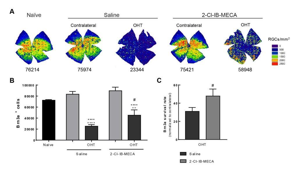
The therapeutic potential of adenosine A3 receptor (A3R) activation was investigated using 2-Cl-IB-MECA, a selective A3R agonist, in an animal model of ocular hypertension (OHT).

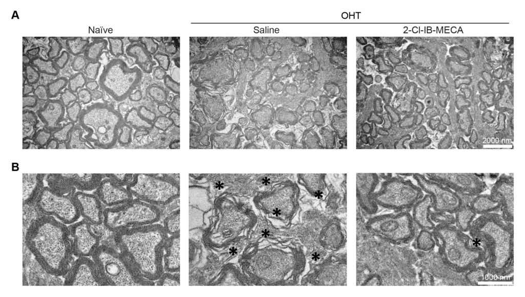
Treatment with 2-Cl-IB-MECA increased RGC survival compared with untreated injured animals. This protective effect was accompanied by reduced impairment of retrograde axonal transport and partial preservation of optic nerve structure, with fewer structural alterations in the axons and their myelin sheaths.

How preclinical models support glaucoma research and the evalua-tion of retinal protection
Figure 3: A3R agonist increases RGC survival in OHT animals. Saline (5 µl) or 2-Cl-IB-MECA (5 µl, 1.2 µM) was administered by intravitreal injection immediately after laser-induced OHT. (A) Representative RGC isodensity maps generated from whole-mount preparations from 2-Cl-IB-MECA and saline-treated OHT, and contralateral retinas immunostained for Brn3a. (B) The number of total Brn3a+ cells per retina was automatically quantified from 5-6 independent retinas. (C) The survival rate of Brn3a+ cells was expressed as the percentage of the ratio between OHT-injured retinas and the contralateral retinas, from 5-6 independent experiments. ++ p < 0.01,++++ p < 0.0001, significantly different from naïve; ****p < 0.0001, significantly different from the contralateral eye; and# p < 0.05, significantly different from saline-treated OHT.
How preclinical models support glaucoma research and the evalua-tion of retinal protection
Figure 4: Treatment with 2-Cl-IB-MECA prevents the structural alterations of optic nerve induced by OHT. Saline (5 µl) or 2-Cl-IB-MECA (5 µl, 1.2 µM) was administered by intravitreal injection immediately after laser-induced OHT. Semi-thin cross sections of naïve, saline-, and 2-Cl-IB-MECAtreated OHT retinas were imaged by transmission electron microscopy. Representative images are depicted at (A) low magnification (scale bar: 2000 nm) and (B) higher magnification (scale bar: 1000 nm). Structural alterations like degenerating axons and myelin disarrangement are observed in OHT animals (indicated by black asterisks).

These findings illustrate the application of established glaucoma models in the evaluation of therapeutic strategies aimed at protecting retinal cells. The protective effects observed with istradefylline were demonstrated in an experimental animal model and do not establish clinical efficacy in patients with glaucoma.

To explore how VectorB2B’s experimental models and retinal assessment capabilities can support your glaucoma research programme, contact our team.

1. Which preclinical models are available for glaucoma research?

VectorB2B has established an in vitro model based on elevated hydrostatic pressure (EHP) and an in vivo model involving transient elevation of intraocular pressure to induce retinal ischemia-reperfusion injury. These models support the investigation of retinal damage and the evaluation of potential therapeutic interventions.

2. How are retinal structure and visual function assessed in animal models?

Retinal structure can be assessed using optical coherence tomography (OCT), while electroretinography (ERG) measures retinal electrical activity. Optomotor response testing provides information about visual function. VectorB2B offers these complementary techniques to support retinal assessment in in vivo glaucoma models.
VectorB2B Articles

Insights and analysis across the life sciences ecosystem, from research and development to regulatory, operational and strategic frameworks.

Find something interesting?
Share it with your peers!

Article topics
Explore other articles