Home Articles Diabetic retinopathy: evaluating therapeutic strategies in preclinical models

Diabetic retinopathy: evaluating therapeutic strategies in preclinical models

Diabetic retinopathy is a major complication of diabetes mellitus and a leading cause of blindness among working-age adults. Preclinical research plays an important role in investigating the mechanisms of retinal damage associated with the disease and evaluating potential therapeutic approaches to limit its progression.

Table of Contents

The research presented here examines retinal alterations using in vitro and in vivo models, including the effects of two pharmacological interventions: SCH 58261 and sitagliptin.

Diabetic retinopathy: background and therapeutic challenges

Diabetic retinopathy involves inflammatory changes and alterations in the neural and microvascular structures of the retina. Glial dysfunction and the breakdown of the blood-retinal barrier (BRB) are among the alterations associated with the disease, with BRB breakdown already evident in its early stages.

Current treatments to limit disease progression include intraocular injections of anti-vascular endothelial growth factor (anti-VEGF) drugs. Laser treatment and surgery are also commonly used.

The evaluation of new therapeutic strategies in animal models of diabetic retinopathy focuses on their ability to:

  • Prevent vascular leakage associated with BRB dysfunction;
  • Control neuroinflammation in the retina;
  • Reduce retinal neuronal cell loss associated with diabetes.

Preclinical models for diabetic retinopathy research at VectorB2B

VectorB2B’s portfolio includes established in vitro and in vivo models of diabetic retinopathy, allowing researchers to investigate retinal alterations associated with diabetes and assess responses to potential therapeutic interventions. These approaches provide complementary experimental settings for studying the cellular and vascular changes involved in the disease.

In vitro models of retinal injury

Retinal cell cultures exposed to elevated glucose concentrations or inflammatory mediators are used to investigate the molecular and cellular mechanisms associated with diabetic retinopathy.

These experimental approaches include primary rat retinal neural cell cultures exposed to high glucose and bovine retinal endothelial cells exposed to inflammatory stimulation. They allow researchers to examine retinal neural cell death and changes in endothelial permeability under controlled experimental conditions.

In vivo models and retinal assessment

VectorB2B has established animal models of both type 1 and type 2 diabetes. These models support the evaluation of blood-retinal barrier (BRB) dysfunction and alterations in retinal structure, including assessments using optical coherence tomography (OCT), and retinal function, assessed by electroretinography (ERG). Additional evaluations focus on neuroinflammation and retinal cell loss, providing information on the neuronal alterations associated with diabetes.

The in vivo findings presented in the following sections were obtained in models of type 1 diabetes, using diabetic mice and rats to investigate retinal damage and responses to pharmacological treatment.

Results from preclinical diabetic retinopathy studies

Studies using animal models of type 1 diabetes have documented blood-retinal barrier (BRB) dysfunction, alterations in retinal structure and increased neuronal cell death. The administration of SCH 58261 and sitagliptin prevented several of these changes, demonstrating the suitability of these experimental models for evaluating potential therapeutic interventions (Aires et al., 2019; Gonçalves et al., 2014).

SCH 58261 and vascular leakage in diabetic mice

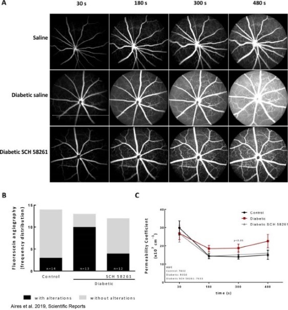
Intravitreal administration of SCH 58261, an adenosine A2A receptor antagonist, reduced vascular leakage in diabetic mice (Aires et al., 2019).

Diabetic retinopathy case study: evaluating retinal damage and therapeutic responses in preclinical models
Fig 1 - Evaluation of vascular leakage with fluorescein angiography assessed in diabetic mice injected intravitreally with SCH 58261. (A) Fluorescein angiography was performed after intraperitoneal injection of 5% fluorescein in control and diabetic non-treated animals or treated with the A2AR antagonist. Images were acquired with the Micron IV (Phoenix Technology) in equivalent time frames. The images represent the distribution of fluorescein across the retina, acquired 30 seconds, 180 seconds, 300 seconds and 480 seconds after injection. (B) The images were classified for the presence or absence of extravasation of fluorescein by 3 independent researchers and the frequency of retinas with and without fluorescein leakage was expressed in percentage of total animals. (C) Permeability coefficient obtained by the quantification of the fluorescence between a major vessel and the interstitial space over time. The area under the curve (AUC) of the permeability coefficient is presented in the graph insert for the different experimental conditions. Permeability coefficient was analyzed with two-way ANOVA using Fisher’s LSD test. (Aires et al. 2019).

Sitagliptin and blood-retinal barrier dysfunction

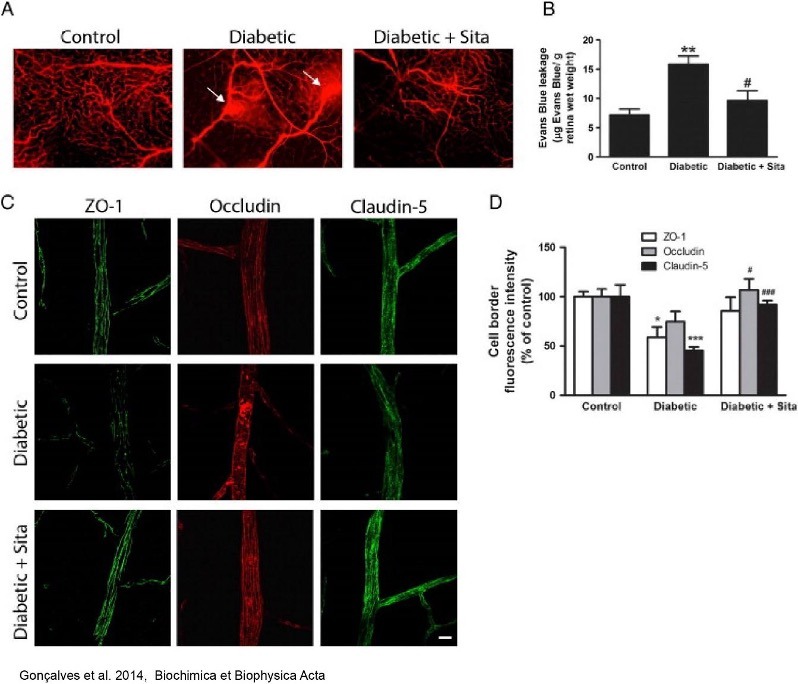
In rats with type 1 diabetes, sitagliptin prevented blood-retinal barrier (BRB) breakdown and alterations in the distribution of tight junction proteins (Gonçalves et al., 2014).

Diabetic retinopathy case study: evaluating retinal damage and therapeutic responses in preclinical models
Fig 2 - Evaluation of the therapeutic response of sitagliptin on BRB permeability and tight junction disassembly triggered by diabetes in rats. (A) Representative images, from 3 animals per group, showing Evans blue fluorescence in the retina. Evans blue, which binds to blood albumin, allows the detection of leakage sites (arrows) in the retinal vessels. Magnification: 100×. (B) Quantitative measurement of BRB permeability by quantification of extravasated Evans blue to retinal parenchyma. Data are presented as μg of Evans blue per retina wet weight (g) and represent the mean ± SEM of 5-6 animals. (C) Sitagliptin prevents the decrease in ZO-1 and claudin-5 immunoreactivity, and the redistribution and accumulation of occludin in rat retinal vessels induced by diabetes. Whole mount preparations of the retinas were imaged by fluorescence confocal microscopy, and images are representative of each group of animals. Magnification 400×. Bar: 20 μm. (D) Quantification of cell border immunostaining for the tight junction proteins (ZO-1, occludin and claudin-5). Data are presented as percentage of control and represent the mean ± SEM of at least 10 fields per retina from 4 animals. *P b 0.05, **P b 0.01 vs. control rats; #P b 0.05, ###P b 0.001 vs. diabetic rats. ANOVA followed by Bonferroni's post hoc test (Gonçalves et al. 2014).

Retinal thickness following SCH 58261 treatment

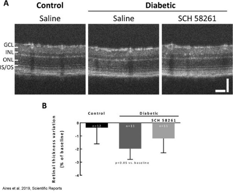
Diabetes was associated with decreased retinal thickness in mice. SCH 58261 administration attenuated this alteration (Aires et al., 2019).

Diabetic retinopathy case study: evaluating retinal damage and therapeutic responses in preclinical models
Fig 3 - Assessment of retinal thickness in diabetic mice injected intravitreally with SCH 58261. Retinal thickness was calculated after layer segmentation in OCT images. (A) Representative OCT images at eight weeks of diabetes. (B) The results were normalized to baseline. Wilcoxon matched-pairs signed rank test. GCL: ganglion cell layer; INL: inner nuclear layer; ONL: outer nuclear layer; IS-OS: photoreceptor inner and outer segments. Scale bars: 50 µm. (Aires et al. 2019).

Microglial reactivity and retinal neuroinflammation

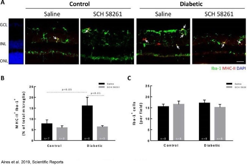
Treatment with SCH 58261 reduced neuroinflammation in the retina of diabetic mice, as investigated through microglial reactivity (Aires et al., 2019).

Diabetic retinopathy case study: evaluating retinal damage and therapeutic responses in preclinical models
Fig 4 - Microglia reactivity in the retina of diabetic mice treated with SCH 58261. (A) Retinal sections were stained with antibodies against Iba-1 (green) and MHC-II (red). Nuclei were stained with DAPI (blue). Representative images are depicted and arrows indicate some MHC-II + Iba-1 + cells found in each condition. (B) Activated microglia (MHC-II + Iba-1 + cells) were counted and normalized to the percentage of total microglial cells (Iba-1 + cells) from 7–9 animals. (C) The number of microglia per retinal section was counted. One-way ANOVA test, followed by Holm-Šídák multiple comparison test. GCL: ganglion cell layer; INL: inner nuclear layer; ONL: outer nuclear layer. Scale bar: 100 µm. (Aires et al. 2019).

SCH 58261 and retinal cell death

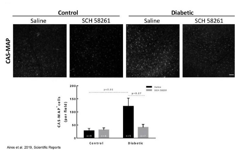
Aires et al. (2019) reported reduced retinal cell death following SCH 58261 treatment. The CAS-MAP assay shown below also indicated fewer cells with active caspases, although this specific comparison did not reach statistical significance.

Diabetic retinopathy case study: evaluating retinal damage and therapeutic responses in preclinical models
Fig 5 - Evaluation of the therapeutic response of SCH 58261 on cell death by apoptosis (CAS-MAP+ cells) triggered by diabetes in mice. The presence of active caspases in the ganglion cell layer was assessed with CASMAP probe (grey). Representative images are depicted. The number of cells CAS-MAP+ was counted from 5–9 independent experiments. One-way ANOVA test, followed by Holm-Šídák multiple comparison test. Scale bar: 100 µm. (Aires et al. 2019)

Sitagliptin and retinal endothelial cell dysfunction

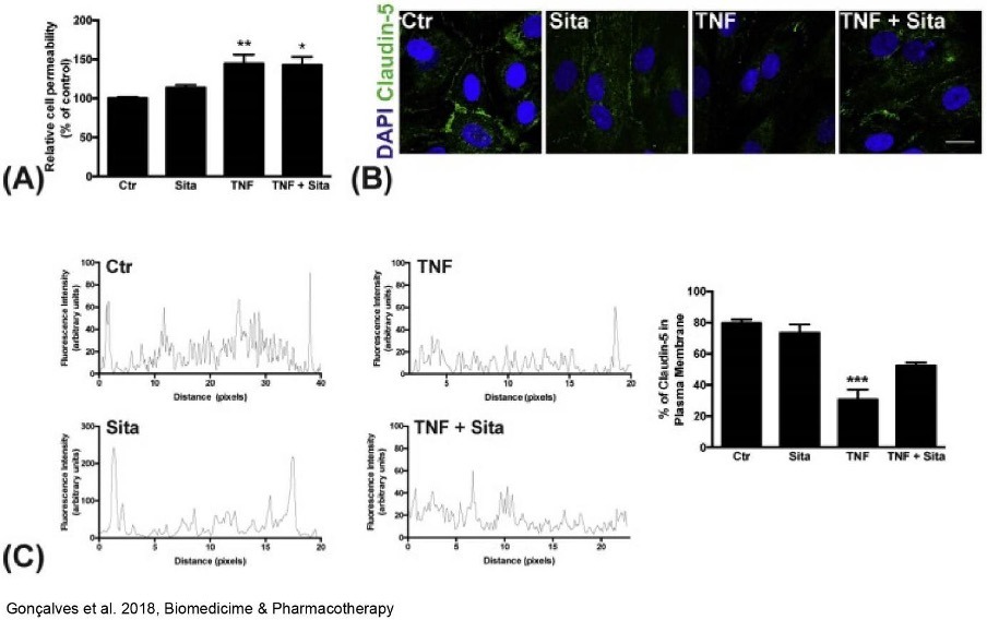
In bovine retinal endothelial cells, tumour necrosis factor alpha (TNF-α), a pro-inflammatory cytokine, increased permeability and altered the distribution of claudin-5. This transmembrane protein is a component of the tight junctions between endothelial cells, which are essential for maintaining blood-retinal barrier (BRB) integrity.

In this experimental setting, sitagliptin did not prevent the TNF-α-induced increase in endothelial permeability (Gonçalves et al., 2018).

Diabetic retinopathy case study: evaluating retinal damage and therapeutic responses in preclinical models
Fig 6 - Impact of sitagliptin on the alterations induced by TNF in the permeability and claudin-5 protein levels of bovine retinal endothelial cells. (A) Bovine retinal EC were grown to confluence on transwell filters and then treated with 5 ng/mL TNF-α, 100 nM sitagliptin or TNF-α plus sitagliptin for 1.5 h. The monolayer permeability to RITC-70 kDa dextran was measured over the following 4 h after the treatments. Sitagliptin was not able to prevent the increase in bovine retinal EC monolayer permeability induced by exposure to TNF-α. The results represent the mean ± SEM of at least 3 independent experiments performed in triplicates. *P < 0.05, **P < 0.01, significantly different from control cells. (B) Confluent bovine retinal EC grown on fibronectin-coated coverslips were treated with TNF-α (5 ng/mL), sitagliptin (100 nM) or TNF-α plus sitagliptin for 6 h. Cells were immunolabeled for claudin-5 and confocal fluorescence images were taken. The images are representative of three independent experiments. Scale bar: 20 μm. (C) Representative plots of intensity profile lines drawn across the interface of adjacent EC were obtained for semi-quantitative analysis of claudin-5 present at the plasma membrane. The results represent the mean ± SEM of n = 4–6. ***P < 0.01, significantly different from control cells. (Gonçalves et al. 2018)

High glucose exposure and retinal neural cell death

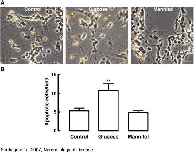
Exposure to elevated glucose concentrations increased apoptotic cell death in primary rat retinal neural cell cultures (Santiago et al., 2007).

Diabetic retinopathy case study: evaluating retinal damage and therapeutic responses in preclinical models
Fig 7 - Elevated glucose increases cell death by apoptosis (TUNEL+ cells) in rat primary retinal neural cell cultures. Rat primary retinal neural cell cultures were incubated in 5 mM glucose (Control), 30 mM glucose (Glucose) or 25 mM mannitol (Mannitol) for 7 days. Cells were labeled by the TUNEL assay. The results represent the mean±SEM of at least three independent experiments and are expressed as the number of TUNEL-positive cells per field. Pictures in A are representative of each condition tested. **p<0.01, significantly different from control; one-way ANOVA followed by Dunnett's post-hoc test. Bar: 20 μm. (Santiago et al. 2007)

Taken together, these findings demonstrate the value of established in vitro and in vivo models for investigating the pathological alterations associated with diabetic retinopathy. They also support their application in the preclinical evaluation of new therapeutic approaches.

To explore how VectorB2B’s experimental models can support your research, contact our team.


Frequently asked questions (FAQ)

1. Why is blood-retinal barrier integrity assessed in diabetic retinopathy research?

Blood-retinal barrier breakdown is an early hallmark of diabetic retinopathy. Preclinical models allow researchers to investigate vascular leakage and alterations in tight junction proteins, helping characterise retinal microvascular dysfunction and evaluate responses to potential therapeutic interventions.

2. How are in vitro and in vivo models used in diabetic retinopathy research?

In vitro models help investigate the molecular and cellular mechanisms associated with diabetic retinopathy, including responses to high glucose and inflammatory mediators. In vivo models allow researchers to assess blood-retinal barrier dysfunction and retinal structural changes, alongside neuroinflammation and neuronal cell loss.

 

Scientific references

  • D. Aires, M. H. Madeira, R. Boia, A. C. Rodrigues-Neves, J. M. Martins, A. F. Ambrósio and A. R. Santiago. Intravitreal injection of adenosine A2A receptor antagonist reduces neuroinflammation, vascular leakage and cell death in the retina of diabetic mice. Scientific Reports 9,17207 (2019). PMID: 31748653.

  • Gonçalves, L. Almeida, A.P. Silva, C. Fontes-Ribeiro, A.F. Ambrósio, A. Cristovão, R. Fernandes. The dipeptidyl peptidase-4 (DPP-4) inhibitor sitagliptin ameliorates retinal endothelial cell dysfunction triggered by inflammation. Biomedicine & Pharmacotherapy 102 (2018) 833–838. https://doi.org/10.1016/j.biopha.2018.03.144. PMID: 29605771.

  • Gonçalves, C. Marques, E. Leal, C. Fontes-Ribeiro, F. Reis, A.F. Ambrósio, R. Fernandes. Dipeptidyl peptidase-IV Inhibition Prevents Blood-Retinal Barrier Breakdown, Inflammation and Neuronal Cell Death in the Retina of Type 1 Diabetic Rats. Biochim Biophys Acta. (2014) Sep;1842(9):1454-63. DOI: 10.1016/j.bbadis.2014.04.013. PMID: 24769045.

  • R. Santiago, A.J. Cristóvão, P.F. Santos, C.M. Carvalho, A.F. Ambrósio. High glucose induces caspase-independent cell death in retinal neural cells. Neurobiol Dis (2007) Mar;25(3):464-72. https://doi.org/10.1016/j.nbd.2006.10.023. PMID: 17239603.

VectorB2B Articles

Insights and analysis across the life sciences ecosystem, from research and development to regulatory, operational and strategic frameworks.

Find something interesting?
Share it with your peers!

Article topics
Explore other articles