
In vivo models in retinal research have become a central experimental tool in ophthalmology research, as they allow the study of retinal dysfunction and degeneration within an integrated biological system. In the context of retinal degenerative diseases, this is particularly relevant. These conditions involve vascular dysfunction, neuronal damage, inflammatory signalling and, in some cases, progressive structural remodelling that cannot be adequately understood through simplified experimental systems alone. |
Table of Contents
Consequently, in vivo models make it possible to investigate:
- How disease develops over time;
- How specific pathological features interact;
- How candidate therapeutic strategies perform in biologically complex settings;
- How disease progression can be monitored through integrated structural and functional endpoints.
In highly specialised development pathways, this capacity directly informs translational decision-making, particularly when the objective is to move from disease understanding towards therapeutic evaluation.
Why in vivo models in retinal research remain central to disease understanding and development
Retinal degenerative diseases continue to present significant scientific and clinical challenges, largely due to the incomplete understanding of the mechanisms that drive their onset and progression. This limitation continues to constrain the translation of current knowledge into sufficiently effective therapeutic strategies across conditions such as diabetic retinopathy, glaucoma and age-related macular degeneration (AMD).
This gap has direct implications for research and development. Early diagnosis remains difficult, reliable biomarkers for disease progression are still needed, and available treatments often address only specific aspects of pathology. Under these conditions, in vivo models in retinal research provide a framework in which disease mechanism can be studied within an integrated biological context, rather than as isolated phenomena.
By enabling the observation of vascular dysfunction, neuronal impairment and inflammatory activity within the same system, in vivo models in retinal research support a more consistent interpretation of disease progression. They also allow therapeutic strategies to be evaluated under conditions that reflect key features of retinal pathology, which is essential when moving from experimental investigation towards clinically relevant development decisions.
The burden and complexity of retinal degenerative diseases
These diseases represent a major source of vision impairment and blindness worldwide, with diabetic retinopathy, glaucoma and age-related macular degeneration among the most prevalent conditions. Vision loss affects the ability to perform everyday tasks, reduces mobility and is associated with broader health risks, including increased likelihood of falls and reduced mental wellbeing.
World Health Organization (WHO) data show that a significant proportion of vision impairment cases could have been prevented or remain insufficiently addressed, reinforcing the importance of early detection and timely intervention. The associated economic impact is equally relevant, with considerable productivity losses linked to visual impairment and long-term care needs. This combination of clinical, social and economic factors places retinal diseases among the most pressing challenges in global health.
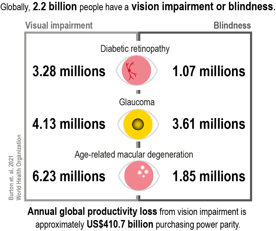
Global burden of retinal degenerative diseases.
This burden is further compounded by the internal complexity of these conditions. Diabetic retinopathy, glaucoma and age-related macular degeneration (AMD) follow distinct pathological pathways, involving different combinations of vascular dysfunction, neuronal degeneration and inflammatory processes. Their progression is often gradual and multifactorial, which complicates both diagnosis and treatment.
In this context, understanding how these mechanisms interact over time remains a central challenge, directly influencing the development of effective therapeutic strategies.
Pathophysiology as a constraint to therapeutic development
The development of effective therapies for retinal degenerative diseases is constrained by the same pathophysiological complexity that defines these conditions. When underlying mechanisms are only partially characterised, therapeutic strategies tend to focus on specific aspects of disease rather than on its full biological scope.
This limitation is evident across major retinal conditions:
- Diabetic retinopathy: blood-retinal barrier breakdown is a defining feature, associated with vascular alterations and a chronic inflammatory environment that contributes to retinal neural dysfunction. This broader characterisation of the disease as neurovascular extends beyond the targets addressed by current therapies;
- Glaucoma: elevated intraocular pressure remains the primary modifiable risk factor and the main focus of treatment. However, disease progression often continues despite pressure control, reflecting the contribution of retinal ganglion cell degeneration and microglia-mediated neuroinflammation;
- AMD: anti-VEGF therapy addresses the neovascular form but requires repeated intravitreal administration and does not cover all aspects of disease progression. The dry form, including geographic atrophy, remains without effective treatment.
These constraints reflect a common challenge: therapeutic development tends to advance around mechanisms that are sufficiently understood and experimentally accessible, while other components of disease progression remain less effectively targeted.H2: Designing in vivo models for retinal diseases
The development of in vivo models in retinal research has played a central role in advancing the understanding of disease mechanisms and in creating the conditions required to evaluate new therapeutic strategies.
Their function is to reproduce specific pathological features in a controlled environment, with model design aligned to the mechanisms under study and adapted to reflect vascular dysfunction, neuronal damage or inflammatory activity depending on the disease and the research objective.
Disease-specific modelling approaches
Across major retinal diseases, modelling strategies are structured around their defining pathological features:
- Diabetic retinopathy: models based on the induction of type 1 and type 2 diabetes, including streptozotocin-induced systems and Goto-Kakizaki animals, reproduce features such as hyperglycaemia, blood-retinal barrier breakdown and microglial activation;
- Glaucoma: experimental systems focus on intraocular pressure elevation through techniques such as laser photocoagulation, episcleral vein cauterisation or microbead injection, alongside pressure-independent models used to study retinal ganglion cell degeneration and neurodegenerative mechanisms;
- AMD: genetic and light-induced models are used to reproduce features of the dry form, while laser-induced choroidal neovascularisation remains the most widely used approach for wet AMD.
Each system reproduces selected aspects of disease, making it possible to analyse specific mechanisms under controlled conditions while preserving physiological context.
At the same time, their scope is inherently selective. They do not capture the full complexity of human retinal disease, and species-specific differences may influence outcomes. For this reason, experimental design must remain closely aligned with study objectives to ensure that the data generated are meaningful for subsequent stages of research and development.
From observation to measurement: monitoring retinal function and structure
The study of retinal degenerative diseases requires the ability to monitor the temporal evolution of disease-related changes. In vivo models in retinal research enable this by supporting the assessment of structural, functional, vascular and visual changes within the same system.
Several techniques used in clinical ophthalmology have been adapted to experimental settings. Fluorescein angiography allows the evaluation of retinal vascular alterations and blood-retinal barrier leakage, while optical coherence tomography provides high-resolution imaging of retinal structure.
Functional assessment is performed through electroretinography, which measures retinal electrical activity, and quantitative optomotor response, which evaluates visual function in animal models.
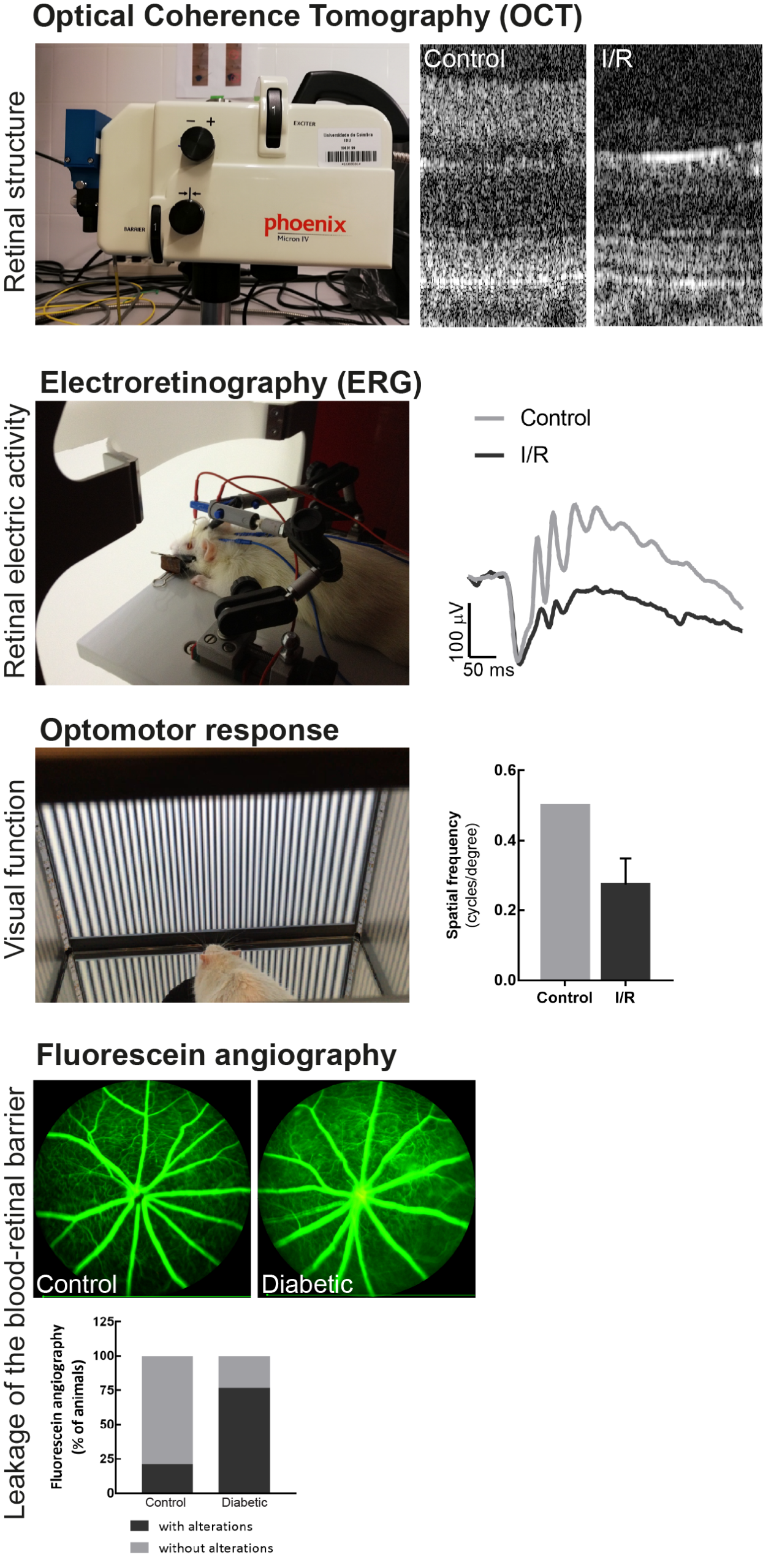
Monitoring retinal disease progression in in vivo models.
Together, these approaches support a more complete characterisation of disease progression, linking structural changes to functional outcomes and strengthening the interpretation of preclinical data in a translational context.
Translational implications of in vivo models in retinal research
In vivo models in retinal research provide a direct link between the study of disease mechanisms and the development of therapeutic strategies. By enabling the controlled investigation of specific pathological features, these models support the identification of relevant targets and the evaluation of interventions under conditions that reflect key aspects of retinal disease.
In diabetic retinopathy, glaucoma and AMD, these systems make it possible to explore mechanisms that extend beyond the primary targets of existing treatments, including neuroinflammatory processes and neuronal degeneration.
From a development standpoint, in vivo models in retinal research contribute to the validation of therapeutic hypotheses and the generation of data that support progression towards clinical evaluation.
VectorB2B supports ophthalmology research programmes through specialised in vivo platforms designed to reflect the complexity of retinal disease, enabling the generation of data aligned with both scientific and translational objectives. If you would like to explore how these capabilities apply to your research programme, contact our team.
Frequently asked questions (FAQ)
1. How do researchers select the most appropriate in vivo model for a specific retinal disease?
Model selection depends on the pathological feature under study, such as vascular dysfunction, neurodegeneration or inflammation. Each system is designed to reproduce specific mechanisms, ensuring alignment between experimental design and study objectives.
2. How early in development should in vivo retinal models be integrated into a research programme?
They become particularly relevant when moving from mechanistic research to therapeutic evaluation. At this stage, they support the assessment of disease progression and treatment response in an integrated biological context.
3. Can retinal models contribute to research beyond ophthalmology, particularly in neurodegenerative diseases?
Yes. The retina shares key characteristics with the central nervous system and can be assessed non-invasively. Structural changes, such as retinal thinning, have been associated with neurodegenerative diseases like Alzheimer’s and Parkinson’s.
VectorB2B Articles
Insights and analysis across the life sciences ecosystem, from research and development to regulatory, operational and strategic frameworks.
Find something interesting?
Share it with your peers!
Article topics
Explore other articles