Xenograft models play a central role in modern oncology research and remain an essential component of preclinical drug development. By enabling human tumour cells or tissues to grow in animal hosts, researchers can evaluate tumour behaviour and therapeutic efficacy in vivo before treatments progress to clinical investigation.
Table of Contents
In highly regulated development environments, these experimental systems provide critical evidence that informs early development decisions.
The use of animal models in cancer research dates back more than a century. The first experimental animal model of cancer was reported in 1918, initiating the systematic use of in vivo systems to investigate tumour biology and therapeutic response. Since then, numerous experimental models have been developed to better reproduce tumour development, progression and treatment response under controlled laboratory conditions.
Among these models, mice have become the preferred species in oncological research due to their short generation intervals, high fecundity, small size and ease of maintenance. Their well-characterised genetic background has enabled the development of specialised strains used in biomedical research, supporting the establishment of both syngeneic and xenograft models that contribute significantly to understanding cancer mechanisms and disease progression.
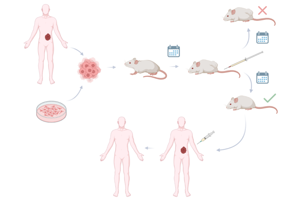
Figure SEQ Figure \* ARABIC 1 – Simplified schematic representation of a xenograft study evaluating compound efficacy, from the animal model to human trials. Created with Biorender.com.
Cell-derived xenograft models
Cell-derived xenograft (CDX) models are widely used in preclinical oncology to investigate tumour growth and evaluate the efficacy of new therapeutic strategies. In this approach, cells from immortalised cancer cell lines derived from patient tumours are implanted into immunodeficient animals, typically mice, allowing tumour development and treatment response to be studied in vivo.
However, CDX models present certain limitations: they rely on cell lines adapted to laboratory culture conditions and therefore do not fully reproduce tumour heterogeneity or the complex interactions between tumours and the immune system observed in patients.
Within this category, two implantation approaches are commonly used in preclinical oncology research:
Ectopic tumour xenograft models
In this method, tumour cells are injected subcutaneously, frequently in the back or hind limb of mice, regardless of the tumour’s original anatomical location. As a result, cultured cells develop in a site different from the one from which they were initially collected.
This implantation approach is technically straightforward, which largely explains its widespread use in oncological research. The subcutaneous location allows tumour growth to be monitored easily and enables researchers to evaluate therapeutic responses in a reproducible experimental setting.
Orthotopic tumour xenograft models
In this approach, tumour cells are transplanted into animals at a site corresponding to their original location. This implantation strategy allows tumours to develop in a biological context that more closely resembles their natural microenvironment, resulting in a more reliable representation of tumour biology and disease progression.
However, unlike subcutaneous ectopic models, tumour growth is usually more difficult to evaluate without sacrificing the animals. In these cases, imaging techniques such as optical imaging, computed tomography or magnetic resonance imaging are commonly used to monitor tumour development. These xenograft systems typically require cell lines expressing fluorescent markers or luciferase, enabling tumour growth to be monitored over time.
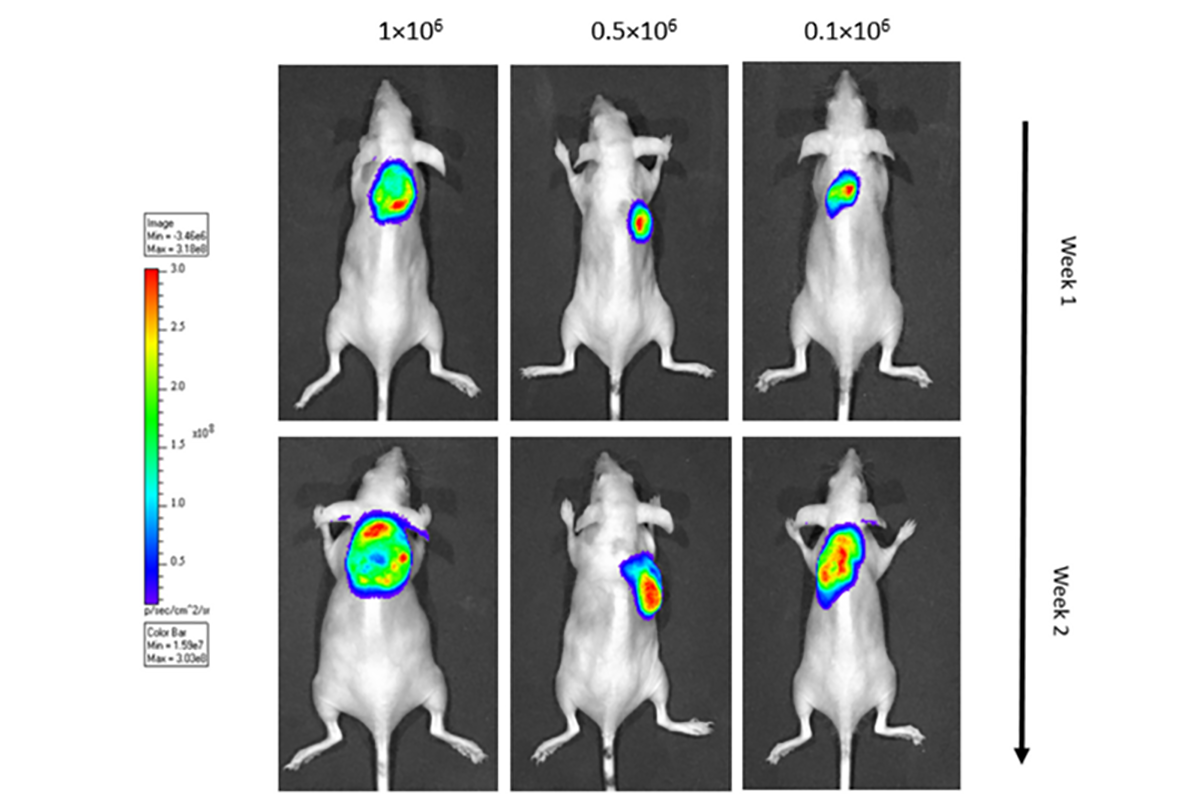
Establishment of a bioluminescent mouse model of canine DLBCL. Bioluminescent imaging of a luciferase-expressing cell line of canine lymphoma injected subcutaneously into immunodeficient mice, via IVIS system. Performed to monitor tumour development.
Advanced xenograft models in cancer research
Although CDX systems remain widely used in preclinical oncology, their reliance on tumour cell lines adapted to in vitro culture conditions limits their ability to reproduce the biological complexity of tumours. These experimental constraints help explain the high failure rate of therapeutic candidates when transitioning from preclinical studies to clinical trials.
Cells that grow outside their natural microenvironment may diverge genetically from the original tumour tissue, altering their biological behaviour and reducing the capacity of these systems to reflect tumour heterogeneity. In response, alternative approaches have been developed to improve the translational relevance of preclinical oncology research:
Patient-derived xenograft (PDX) models
This approach involves the inoculation of tumour tissue obtained directly from patients into immunodeficient mice. Because the tumour material is not first adapted to in vitro culture conditions, these systems preserve many of the genetic and histological characteristics of the original tumour.
It enables researchers to study tumour heterogeneity and explore potential correlations between tumour characteristics and disease progression. However, the establishment of PDX systems presents practical challenges. Tumour samples must be transported rapidly from the operating room to the laboratory, and histological analysis is typically performed prior to inoculation.
Even under these conditions, engraftment rates remain limited, with reported implantation rates of approximately 25%.
Humanized mouse models
Conventional xenograft models rely on immunocompromised animals, which restricts their ability to reproduce interactions between tumour cells and the immune system. Humanized mouse models were developed to address this limitation.
In these systems, immunodeficient mice are engrafted with human haematopoietic stem cells, enabling the development of components of a human immune system. This experimental framework allows researchers to investigate immune–tumour interactions and evaluate therapeutic strategies such as immunotherapies within a more representative biological context.
Immunocompromised animal models used in xenograft research
Xenograft models rely on immunocompromised animal systems that allow tumour cells from another species to engraft and proliferate. Over time, several strains of immunodeficient animals have been developed to support oncology research and other biomedical applications.
The first immunodeficient mouse model was described in 1966 with the identification of the nude mouse, which lacks functional T lymphocytes due to abnormal thymus development. Subsequent studies led to the discovery of mice with severe combined immunodeficiency (SCID), characterised by defects affecting both T and B lymphocyte development.
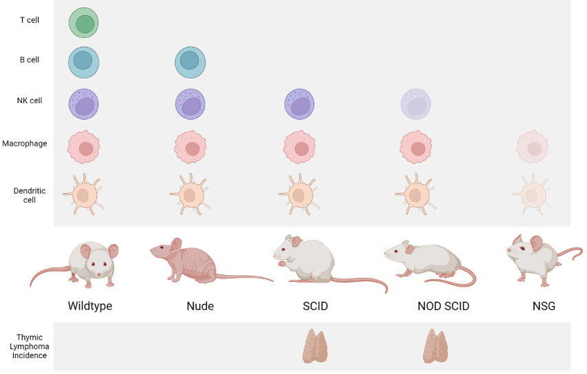
Schematic representation of commonly used immunodeficient mouse models. Created with Biorender.com
Further genetic modifications have resulted in additional strains with deeper levels of immunodeficiency. Among these, NOD/SCID mice and NOD/SCID rgnull mice are widely used in xenograft research due to their increased capacity to support human cell engraftment.
Several genes play a critical role in determining the level of immunodeficiency observed in these animal models.
Key genes associated with immunodeficiency in animal models
| Gene | Characteristics |
|---|---|
|
Forkhead Box N1 (FOXN1) |
Functional deficiency of FOXN1 leads to a nude SCID animal, with retarded growth and lower fertility. The thymus is absent at birth in these mice, and very few lymphocytes occur in the spleen and lymph nodes. |
|
DNA-Dependent Protein Kinase Catalytic Subunit (PRKDC) |
Several mammal species (including humans) with defective PRKDC genes have an inhibition of lymphocyte development, resulting in SCID with an absence of functional T and B cells. |
|
Interleukin 2 Receptor Subunit Gamma (IL2RG) |
The IL2RG is located on the X chromosome. This gene is essential for interleukin signaling pathways, which regulate T-cell differentiation and peripheral tolerance, increase the cytolytic activity of NK cells and regulate B-cell differentiation. |
|
Recombination Activating Gene 1 and 2 (RAG1 and RAG2) |
Defects in these genes lead to a severe block or deficient generation of T and B cells. SCID resulting from defects in RAG1 and RAG2 is characterized by severe depletion in mature T and B cell numbers, whereas NK cells are present in normal numbers. |
|
Janus Kinase 3 (JAK3) |
JAK3 deficiency is associated with the absence of T lymphocytes, NK cells and the presence of non-functional B lymphocytes. |
|
Artemis (DCLRE1C) |
Artemis deficiency is an autosomal recessive disorder that affects the mechanism of recombination of the T cell receptor and of B cell receptor complexes. |
|
Beta-2-Microglobulin (B2M) and Perforin 1 |
B2M is related to the development of cytotoxic T cells and NK cell function. |
|
Adenosine Deaminase (ADA) and Adenylate Kinase 2 (AK2) |
ADA deficiency can cause comprehensive lymphocyte apoptosis, leading to a SCID with severe T, NK and B lymphocytopenia. AK2 deficiency, also known as reticular dysgenesis, displays SCID with absence of T, B and NK cells. |
|
Coronin-1A (CORO1A) |
CORO1A deficient mice have reduced peripheral T cells due to increased apoptosis, causing T-/B+/NK+SCID. |
Beyond mice, other animal species may also be used in cancer research. Cases of severe combined immunodeficiency have also been reported in rabbits, dogs and pigs, expanding the range of experimental systems available for cancer research.
Future perspectives for preclinical cancer models
In oncology research, xenograft models and other advanced preclinical platforms are increasingly refined to improve how accurately experimental systems reproduce the biological characteristics of human tumours and their microenvironment. At the same time, the limitations of existing in vivo systems have encouraged the exploration of complementary approaches capable of providing additional insights into tumour behaviour.
The continuous refinement of preclinical platforms, together with advances in molecular biology and imaging technologies, is expected to strengthen the contribution of preclinical research to the development of safer and more effective cancer therapies.
In oncology drug development, these experimental systems generate critical evidence supporting the transition from early research to clinical investigation. Xenograft models remain central to this process, enabling researchers to evaluate therapeutic efficacy and identify potential risks before new treatments reach human trials.
VectorB2B supports biotechnology and pharmaceutical organisations across the drug development pathway, from early discovery studies to clinical development. Through specialised preclinical platforms, research teams can generate robust data that support oncology programmes. If you would like to explore how advanced preclinical models can support your oncology research programme, explore our services.
Frequently asked questions (FAQ)
1. What factors influence the success of tumour engraftment in xenograft models?
In these approaches, engraftment success depends on the biological characteristics of the tumour tissue, the degree of immunodeficiency of the host animal and the procedures used during tumour inoculation.
2. How is tumour growth monitored in xenograft studies?
In many xenograft models, tumour growth can be measured directly in subcutaneous studies. In other experimental settings, imaging techniques such as optical imaging, CT or MRI are used to monitor tumour development.
3. Why are xenograft models important in oncology research?
They provide an experimental framework for studying tumour behaviour and evaluating therapeutic strategies before clinical investigation. Within preclinical oncology research, xenograft models generate evidence that supports the assessment of treatment response and disease progression.
VectorB2B Articles
Insights and analysis across the life sciences ecosystem, from research and development to regulatory, operational and strategic frameworks.
Find something interesting?
Share it with your peers!
Article topics
Explore other articles